Beranda
/ Anatomy Of Back Of Neck And Head : Anatomy of the Head and Neck | Human Anatomy | Medical ... : 1 also, some people are more prone to headaches than others.
Anatomy Of Back Of Neck And Head : Anatomy of the Head and Neck | Human Anatomy | Medical ... : 1 also, some people are more prone to headaches than others.
Insurance Gas/Electricity Loans Mortgage Attorney Lawyer Donate Conference Call Degree Credit Treatment Software Classes Recovery Trading Rehab Hosting Transfer Cord Blood Claim compensation mesothelioma mesothelioma attorney Houston car accident lawyer moreno valley can you sue a doctor for wrong diagnosis doctorate in security top online doctoral programs in business educational leadership doctoral programs online car accident doctor atlanta car accident doctor atlanta accident attorney rancho Cucamonga truck accident attorney san Antonio ONLINE BUSINESS DEGREE PROGRAMS ACCREDITED online accredited psychology degree masters degree in human resources online public administration masters degree online bitcoin merchant account bitcoin merchant services compare car insurance auto insurance troy mi seo explanation digital marketing degree floridaseo company fitness showrooms stamfordct how to work more efficiently seowordpress tips meaning of seo what is an seo what does an seo do what seo stands for best seotips google seo advice seo steps, The secure cloud-based platform for smart service delivery. Safelink is used by legal, professional and financial services to protect sensitive information, accelerate business processes and increase productivity. Use Safelink to collaborate securely with clients, colleagues and external parties. Safelink has a menu of workspace types with advanced features for dispute resolution, running deals and customised client portal creation. All data is encrypted (at rest and in transit and you retain your own encryption keys. Our titan security framework ensures your data is secure and you even have the option to choose your own data location from Channel Islands, London (UK), Dublin (EU), Australia.
Anatomy Of Back Of Neck And Head : Anatomy of the Head and Neck | Human Anatomy | Medical ... : 1 also, some people are more prone to headaches than others.. The mayo clinic says that these factors can be chemical activity in your brain, the nerves of blood vessels surrounding your skull, or the muscles at the back of the head and neck. It controls flexion, lateral flexion, and rotation of the vertebral column, and maintains the lumbar curve. These muscles can extend the head, laterally flex it, and rotate it ((figure)). See anatomy of the head and neck stock video clips. Ct scan of head and neck :
Occipital neuralgia is caused due to irritation or injury to the occipital nerve. The superficial lymph nodes of the head and neck receive lymph from the scalp, face and neck. Radiological anatomy of the head and neck on a ct in axial, coronal, and sagittal sections, and on a 3d images The neck muscles, including the sternocleidomastoid and the trapezius, are responsible for the gross motor movement in the muscular system of the head and neck. Neck muscles are bodies of tissue that produce motion in the neck when stimulated.
CLASS BLOG: BIO 202 Arteries and Veins KEY from 1.bp.blogspot.com 1 also, some people are more prone to headaches than others. Think of it like a jigsaw puzzle, all the pieces fit in together and are required to get the full picture as to how it works. The superficial lymph nodes of the head and neck receive lymph from the scalp, face and neck. It also includes the veins that return deoxygenated blood from these organs to the heart. The external jugular veins descend on either side of the neck, passing over the sternomastoid muscles and beneath the platysma. The posterior muscles of the neck are primarily concerned with head movements, like extension. The muscles of the neck run from the base of the skull to the upper back and work together to bend the head and. The neck is connected to the upper back through a series of seven vertebral segments.
Muscles of the neck and back
They empty into the right and left subclavian veins in the base of the neck. The skull can be further subdivided into: Included in this review are the cervica. The back muscles stabilize and move the vertebral column, and are grouped according to the lengths and direction of the fascicles. The skeletal section of the head and neck forms the top part of the axial skeleton and is made up of the skull, hyoid bone, auditory ossicles, and cervical spine. From the sides and the back of the neck, the splenius capitis inserts onto the head region, and the splenius cervicis extends onto the cervical region. 1 also, some people are more prone to headaches than others. It controls flexion, lateral flexion, and rotation of the vertebral column, and maintains the lumbar curve. Internal and external carotid arteries The external jugular veins descend on either side of the neck, passing over the sternomastoid muscles and beneath the platysma. The top of the cervical spine connects to the skull, and the bottom connects to the upper back at about shoulder level. The large, complex muscles of the neck and back move the head, shoulders, and vertebral column. Their afferent vessels drain the occipital region of the scalp, while their efferents pass to the superior deep cervical glands.
The head and neck are more than just features used to identify your friends or relatives. The neck muscles, including the sternocleidomastoid and the trapezius, are responsible for the gross motor movement in the muscular system of the head and neck. Each nerve provides sensation to a specific area of the body called a dermatome. The skull can be further subdivided into: The nerves of the head include the sympathetic and parasympathetic innervation to the head and neck, as well as the three branches of the trigeminal nerve:
Anatomy of the back: Spine and back muscles | Kenhub from thumbor.kenhub.com They ultimately drain into the deep lymph nodes. Internal and external carotid arteries These muscles can extend the head, laterally flex it, and rotate it ((figure)). Finger , wrist , head , neck , thigh , knee , leg , shoulder , arm , forearm , thorax , ankle. Ct scan of head and neck : Their afferent vessels drain the occipital region of the scalp, while their efferents pass to the superior deep cervical glands. It also includes the veins that return deoxygenated blood from these organs to the heart. Knowing what causes headaches can help you know how to.
The erector spinae group forms the majority of the muscle mass of the back and it is the primary extensor of the vertebral column.
The top of the cervical spine connects to the skull, and the bottom connects to the upper back at about shoulder level. The pain in the back of your head occurs when one or a combination of factors affect your brain. The head rests on the top part of the vertebral column, with the skull joining at c1 (the first cervical vertebra known as the atlas). The muscles of the neck run from the base of the skull to the upper back and work together to bend the head and. The superficial lymph nodes of the head and neck receive lymph from the scalp, face and neck. Radiological anatomy of the head and neck on a ct in axial, coronal, and sagittal sections, and on a 3d images Muscles of the neck and back The skull can be further subdivided into: An area called the occiput. The neurocranium (cranial vault) and the viscerocranium (facial skeleton). These diverse tasks require both strong, forceful movements and some of the fastest, finest, and most delicate adjustments in the entire human body. The back muscles stabilize and move the vertebral column, and are grouped according to the lengths and direction of the fascicles. From the sides and the back of the neck, the splenius capitis inserts onto the head region, and the splenius cervicis extends onto the cervical region.
They are arranged in a ring shape; The posterior muscles of the neck are primarily concerned with head movements, like extension. The splenius muscles originate at the midline and run laterally and superiorly to their insertions. Neck muscles are bodies of tissue that produce motion in the neck when stimulated. The neck muscles, including the sternocleidomastoid and the trapezius, are responsible for the gross motor movement in the muscular system of the head and neck.
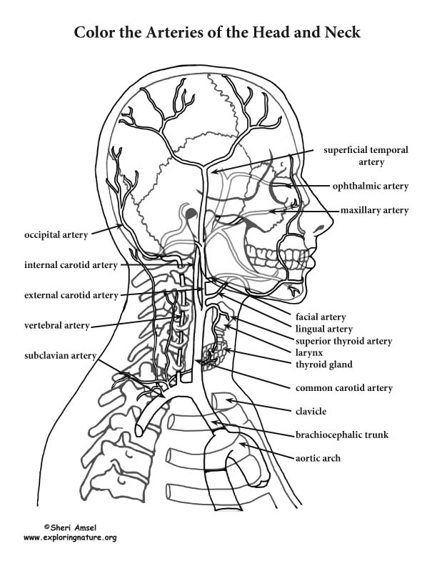
Arteries of the Head and Neck Coloring Page from www.exploringnature.org Knowing what causes headaches can help you know how to. The most important arteries and nerves of the head and neck are the following: Neck muscles are bodies of tissue that produce motion in the neck when stimulated. Labeled anatomy chart of neck and back muscles on white background labeled human anatomy diagram of man's neck and back muscles from a posterior view on a white background. From the sides and the back of the neck, the splenius capitis inserts onto the head region, and the splenius cervicis extends onto the cervical region. Neck anatomy nerves picture there are 8 spinal nerves that originate from the cervical spine. The head rests on the top part of the vertebral column, with the skull joining at c1 (the first cervical vertebra known as the atlas). The large, complex muscles of the neck and back move the head, shoulders, and vertebral column.
The large, complex muscles of the neck and back move the head, shoulders, and vertebral column.
The nerves of the head include the sympathetic and parasympathetic innervation to the head and neck, as well as the three branches of the trigeminal nerve: The splenius muscles originate at the midline and run laterally and superiorly to their insertions. It consists of two major parts: The mayo clinic says that these factors can be chemical activity in your brain, the nerves of blood vessels surrounding your skull, or the muscles at the back of the head and neck. The skull is a strong, bony capsule that rests on the neck and encloses the brain. Neck muscles are bodies of tissue that produce motion in the neck when stimulated. The neurocranium (cranial vault) and the viscerocranium (facial skeleton). These diverse tasks require both strong, forceful movements and some of the fastest, finest, and most delicate adjustments in the entire human body. The posterior muscles of the neck are primarily concerned with head movements, like extension. The neck is connected to the upper back through a series of seven vertebral segments. Occipital neuralgia is caused due to irritation or injury to the occipital nerve. It is made up of bones, discs, muscles, ligaments, nerves and tendons. The skeletal section of the head and neck forms the top part of the axial skeleton and is made up of the skull, hyoid bone, auditory ossicles, and cervical spine.
The occipital glands (lymphoglandulæ occipitales), one to three in nu ber, are placed on the back of the head close to the margin of the trapezius and resting on the insertion of the semispinalis capitis anatomy of back of neck. The mayo clinic says that these factors can be chemical activity in your brain, the nerves of blood vessels surrounding your skull, or the muscles at the back of the head and neck.